


An international research team has achieved a major breakthrough in structural biology by determining a high-resolution protein structure from a single crystal grown inside a living cell. The achievement was made possible by a newly developed method called IncelluloED.
The research team was led by Vitaly Polovinkin from the ELI Beamlines Facility of the Extreme Light Infrastructure ERIC in Prague and by Lars Redecke from the Institute of Biochemistry at the University of Lübeck and the German Electron Synchrotron (DESY), Hamburg. The collaboration also included scientists from the Institute of Molecular Genetics of the Czech Academy of Sciences (CAS), Prague, the University of South Bohemia in České Budějovice, the Biology Centre of CAS in České Budějovice, the European XFEL in Hamburg, Germany, and the Department of Cell and Molecular Biology at Uppsala University, Sweden.
The method, IncelluloED, combines intracellular protein crystallization with in situ three-dimensional electron diffraction. “Using widely available cryo-electron microscopy tools, IncelluloED enables high-resolution structural analysis directly inside cells, eliminating the need for protein purification or the growth of large crystals outside their native environment“, explained Vitaly Polovinkin.
Intracellular crystallization has emerged as a promising strategy in structural biology, but existing approaches face major limitations. In 2024, the InCellCryst pipeline was introduced by researchers from the University of Lübeck (https://doi.org/10.1038/s41467-024-45985-7) for studying intracellular crystals using serial X-ray crystallography. “However, serial crystallography requires the exposure of tens of thousands of cells, which prevents high-resolution studies on proteins that crystallize only in a few cells”, said Lars Redecke. IncelluloED overcomes this barrier by enabling structure determination from a single crystal within a single cell.
The researchers demonstrated the power of the method using a microcrystal of the HEX-1 protein from Magnaporthe grisea, grown in an insect cell. “IncelluloED achieved a structure at 1.9 Å resolution from a crystal volume of approximately 1.6 µm³, compared with a 1.8 Å structure obtained by serial X-ray crystallography from a combined crystal volume exceeding 11 million µm³. Thus, IncelluloED offers a multimillion-fold reduction in the sample volume required for high-resolution structure determination.
Beyond its immediate performance, IncelluloED opens new avenues for systematic studies of intracellular crystallization, including determining how frequently proteins crystallize in cells, how many intracellular crystals diffract to high resolution, and how well-ordered regions can be identified within a crystal lamella. Future developments, such as introducing small-molecule cofactors into host cells, may further expand the method’s potential for structure-based drug discovery.
Despite the apparent complexity of the workflow, the pipeline proved to be robust: no crystals were lost during the study, and all diffracted to high resolution during tests. By requiring only a single crystal inside a single cell and using standard cryo-EM platforms, IncelluloED brings high-resolution structural biology into conventional laboratory settings. The method represents a significant step toward a new paradigm in the field, the realization of a single-cell structural biology laboratory.
Reference: Štěpánka Bílá, Dominik Pinkas, Krishna Khakurel, Juliane Boger, Tomáš Bílý, Janos Hajdu, Zdeněk Franta, Iñaki de Diego Martinez, Roman Tuma, Lars Redecke and Vitaly Polovinkin, Single-Cell Structural Biology with Intracellular Electron Crystallography, Nature Communications 17, 2109 (2026).
Single-cell structural biology with intracellular electron crystallography | Nature Communications
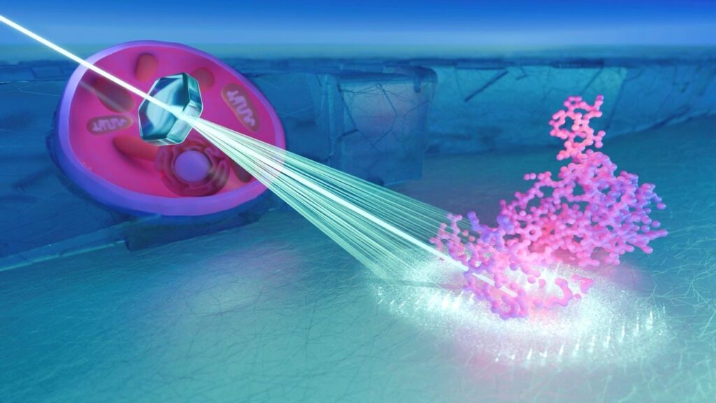
Single-Cell Structural Biology with Intracellular Electron Crystallography. IncelluloED combines intracellular crystallization with 3D electron diffraction as a step toward establishing a “single-cell structural biology laboratory.” Compared with synchrotron-based X-ray techniques, the new method offers a multimillion-fold reduction in the sample volume required for high-resolution 3D structural studies.
Illustration: Lucas J. Martin, Max Planck Institute of Biophysics, Frankfurt am Main, Germany


