The latest specifications of individual scientific instruments can be found on the ELI User Portal.
Secondary sources in E1
High harmonic generation (HHG): This beam line delivers a coherent collimated beam of photons with energies in the range 10 eV–120 eV.
Plasma X-ray source (PXS): This is an incoherent source of hard X-ray radiation.
 HHG Beam LineRead more |
 Plasma X-Ray Source (PXS)Read more |
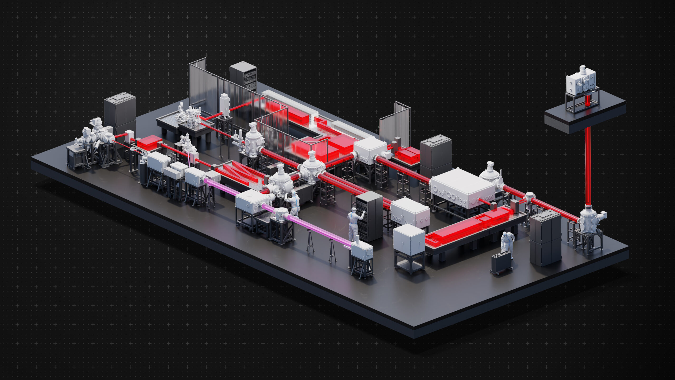
Scientific stations in E1
MAC: a Multi-purpose chamber for AMO (Atomic, molecular, and optical) sciences and CDI (Coherent Diffractive Imaging).
ELIps: A scientific station for time-resolved spectroscopy ellipsometry in the optical range and XUV materials science.
Hard X-ray end-station: TREX; a modular station for Time Resolved Experiemnts (scattering, diffraction, spectroscopy, pulse radiolysis and imaging) with X-rays.
Ultrafast optical spectroscopy including setups for stimulated Raman scattering, transient optical absorption, IR (1D and 2D) and pulse-shaping and coherent control.
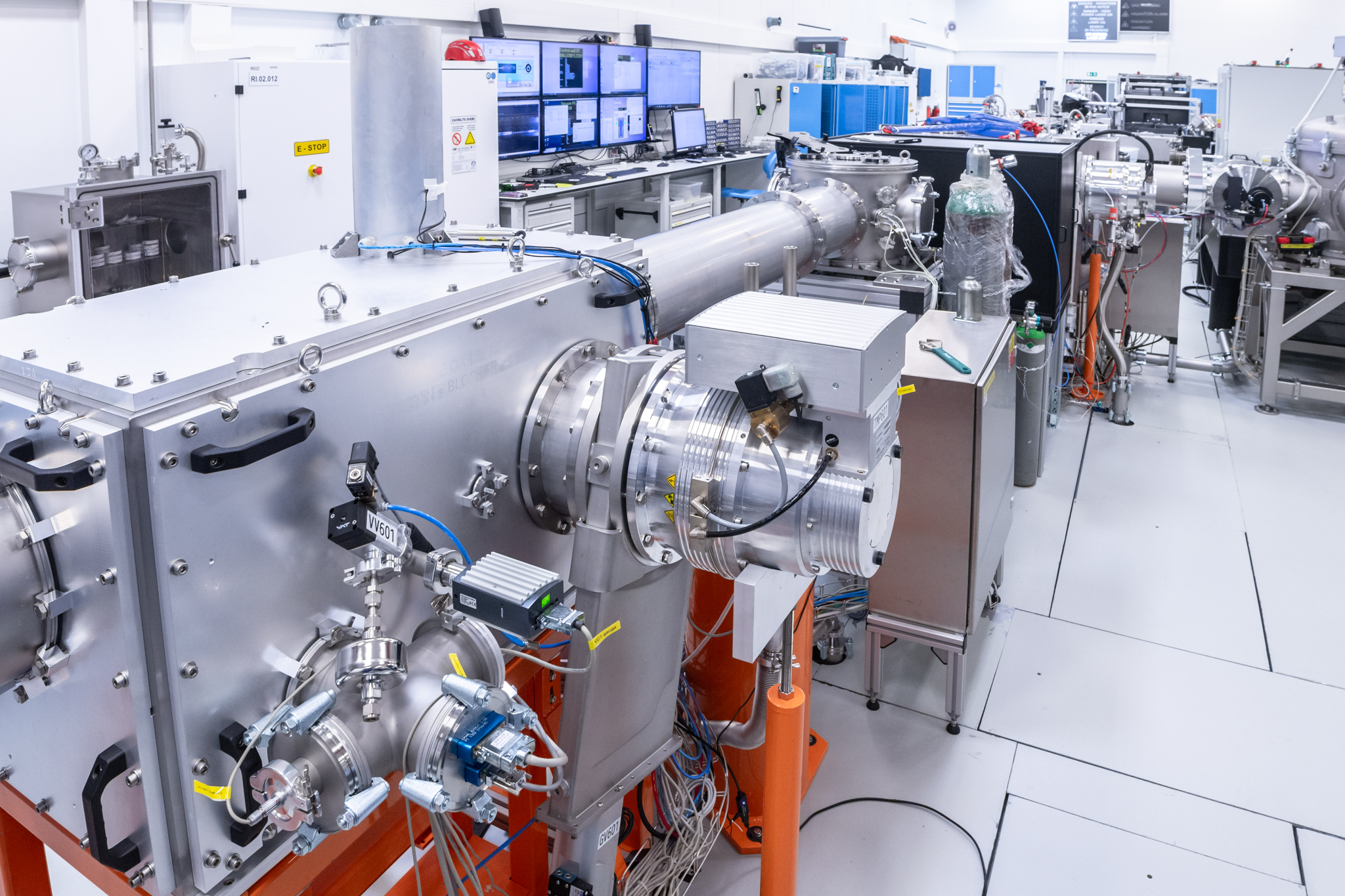
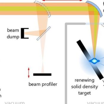
The PXS beam line
operates with the in-house developed L1 Allegra. X-ray radiation is in the spectral range of 3–77 keV. Using an off-axis parabola, the laser beam is focused onto the continuously restoring solid-density target, enabling long-term 1 kHz operation of the beam line. The beam delivery chamber and the plasma interaction chamber are separated by an anti-reflection (AR)-coated quartz window to suppress contamination of the beam transport system by debris. Several diagnostics will be implemented in order to monitor the driving laser beam and the output X-ray radiation:
- An imaging system to monitor the position and spatial profile of the visible emission from the plasma plume (integral part of PXS)
- A focal spot imaging system to monitor the focusing of the attenuated driving laser beam (integral part of PXS)
- An X-ray spectrometer, consisting of an X-ray photodiode, an amplifier, and a digital pulse processor emission spectra measurement (integral part of PXS)
- On-shot spectrometer and ultra-fast photodiode to analyze the shot-to-shot stability and determine the flux as an online monitoring system (additional monitoring system)

The X-ray output port has three output Beryllium windows: two for user end stations and one for the X-ray emission monitor. Following end stations are deployed at the PXS beam line:
1. Goniometer-based diffractometer including a Cu-Ka X-ray tube to provide CW X-ray pump beam and additional X-ray analysis
2. Von Hamos X-ray absorption/emission spectrometer
Both end stations are complemented by single-photon counting (Dectris EIGER X 1M) and charge integrating (Princeton MTE 2048B, Andor iKON B-DD) X-ray detectors as well as scintillator-based photodiode arrays (Hamamatsu S-11866-128G-2).