


TREX: Hard X-ray science, diffraction and scattering, spectroscopy, pulse radiolysis and imaging
Staff:
Borislav Angelov
Martin Precek
Vitaly Polovinkin
Krishna Khakurel
Anna Zymaková
Iuliia Baranova (PhD student)
Brief description of the available set up:
The TREX instrument for X-ray science is developed to be used with a laser driven pulse Plasma X-ray Source (PXS). This source is under commissioning but is still not available for user experiments. In the mean time, two sealed X-ray tube sources (one with a Cu anode for diffraction applications and one with a Mo anode for spectroscopy applications) are available for methods development and early experiments.
X-ray diffraction and scattering
The X-ray diffractometer is based on a custom modification of a commercial STOE STADIVARI goniometer. It has the same functionality as the commercial analog plus in addition an extended range for the detector movement going up to 400 mm sample to detector distance. The main module of the diffractometer is the so called Euler cradle goniometer, which is capable of simultaneously rotating the investigated sample at 360 degree and at the same time to position the X-ray detector at desired angle and distance from the sample. It comes with a computer controlled video microscope and dimmable led light. The cryo cooling for the fragile biological sample is implemented via a commercial cryo stream cooler from Oxford Cryosystems. The recording of the diffracted and scattered X-ray photons is accomplished by a single photon counting hybrid pixel detector model Eiger X 1M from the Dectris company. Presently available X-ray source for the diffraction station is an X-ray microfocus sealed tube form AXO Dresden with Montel optics and JJ X-ray pinhole collimation.
The instrument for hard X-ray science is outlined in the figure below
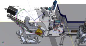
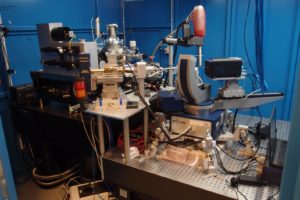
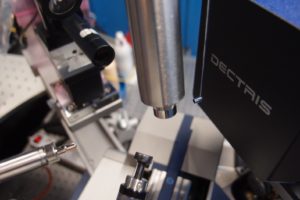
Fig. Top: Hard X-ray experimental station as designed, including the laser driven Plasma X-ray source, a complementary sealed tube Cu-anode X-ray source, diffractometer and von Hamos spectrometer for absorption spectroscopy. Middle: Diffraction station as built (presently available for users with the complementary sealed tube X-ray source). Bottom: detail of the diffractometer sample environment when optimized for protein crystallography with the cryostream cryocooler.
Technical Data: Diffraction station run with conventional sealed tube X-ray source
| Sealed tube X-ray beam parameters | |
| Flux on the sample | 10^8 ph/sec |
| Beam size | 145 micrometers |
| Beam divergence | 4.8 mrad |
| Beam polarization | 40% |
| Wavelength | CuKa 1.54 Angstroms |
| Detector parameters | |
| Pixel size [µm2] | 75 x 75 |
| Sensitive area (width x height) [mm2] | 77.2 x 79.9 |
| Total number of pixels | 1030 x 1065 = 1,096,950 |
| Maximum frame rate [Hz] | 3000 |
| Frame dead time | 3 μs |
| Point-spread function | 1 pixel |
| Sensor thickness [μm] | 450 |
| Threshold energy [keV] | 2.7-18 |
| Maximum count rate [phts/s/mm2] | 5×10^8 |
| Counter bit depth [bit] | 12 |
| Image bit depth [bit] | 16 or 32 |
| Photon processing time per pixel | 180 ns |
| Data format | HDF5 |
| Sample to detector distance | 40-400 mm |
X-ray spectroscopy
The X-ray spectroscopy station is based on the von Hamos design [J. Szlachetko et al., Rev.
Sci. Instrum. 83, 103105 (2012)]. A number of gratings are available and the instrument has the necessary flexibility to cover an energy range of at least 4 up to 12 keV. Initial design of the set up is optimized for absorption spectroscopy but emission spectroscopy can also be supported.
The detector is a custom designed CCD from Andor, based on Newton model, and allows greater acceptance angles. This results in a widened field of view for broader energy window observable within a single experimental acquisition. The detector matrix is of a deep-depleted front-illuminated type that allows preventing charge splitting effects. Beryllium window filters out background illumination efficiently. Presently available X-ray source for the spectroscopy station is a Molybdenum anode X-ray microfocus sealed tube from XOS with policapillary focusing optic. On top of that, an X-ray water-jet source is being under development and optimization to fit the current setup. Generating a broad continuous energy spectrum (Bremsstrahlung emission is a dominant mechanism), the source will serve for X-ray spectroscopy applications, coming soon.
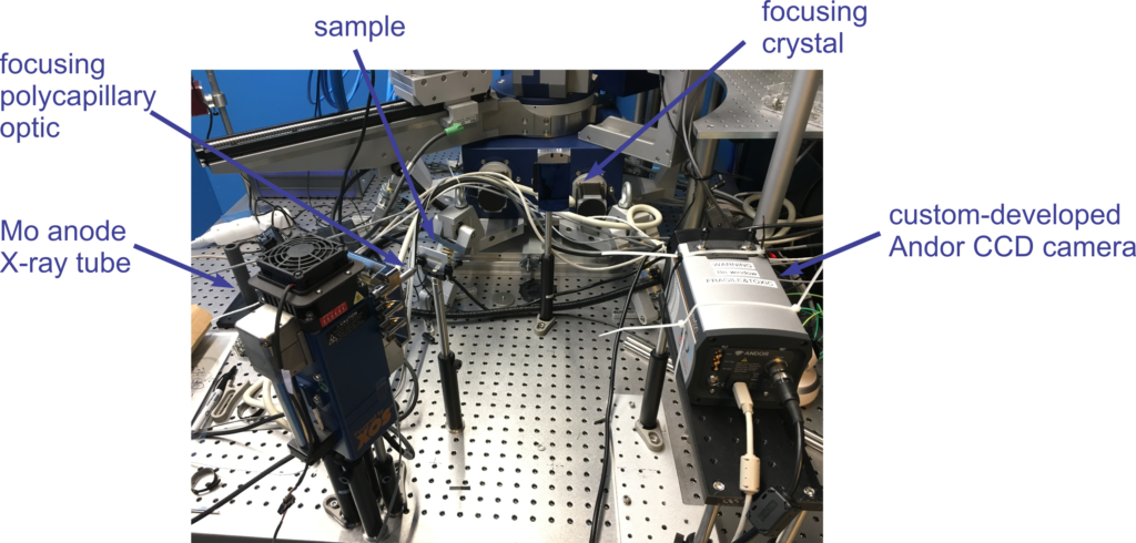
Fig: X-ray spectroscopy station (in absorption geometry) as built in a dedicated X-ray hutch
Technical Data: Spectroscopy station run with conventional XOS X-ray tube
| Sealed tube X-ray beam parameters | |
| Generated flux | 6.1 x 10⁸ ph/sec @ 50 W power |
| Beam size | 68.6 µm (FWHM) @ Mo Kα (17.4 keV) |
| Measured output beam divergence | 3.80 |
| Focal length | 30 mm |
| Detector parameters | |
| Pixel size [µm2] | 26 x 26 |
| Sensor size (width x height) | 1024 x 256 pixels |
| Full well capacity | Higher than 200000 electrons |
| Linearity | Better than 99% |
| Sensor QE | Better than 35% at 8 keV |
| Maximum spectra rate [spectra/sec] | 1612 |
| Dark current [electrons/pixel/sec] | 0.0001 |
Pulse Radiolysis
coming soon
Water jet X-ray source
In collaboration with Lund University in Sweden we are implementing a laser-driven water jet X-ray source for user applications. The source is designed and developed by Dr. Jens Uhlig at the division of Chemical Physics at Lund University (http://www.chemphys.lu.se/people/senior-staff/jens-uhlig/). It is a custom adaptation of the source already successfully described and used in ultrafast X-ray science [1], [2]. The main adaptations serve to prepare the source for using higher laser pulse energies. Following commissioning the source will be made available for the user community.
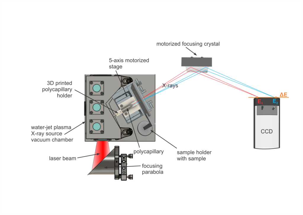
Fig 1: Schematic set up for X-ray spectroscopy using the Lund water jet X-ray source and a von Hamos spectrometer.
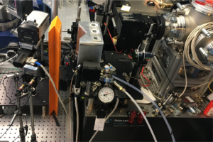
Fig 2: X-ray spectroscopy end station with Water Jet Plasma X-ray Source at the ELI Beamlines facility
Contact person at ELI Beamlines for the Water Jet Source project is Anna Zymakova, anna.zymakova@eli-beams.eu
References:
[1] Laser plasma x-ray source for ultrafast time-resolved x-ray absorption spectroscopy
Miaja-Avila, et al. Structural Dynamics 2, 024301 (2015); https://doi.org/10.1063/1.4913585
[2] Table-Top Ultrafast X-Ray Microcalorimeter Spectrometry for Molecular Structure
Uhlig, et al. Phys. Rev. Lett. 110, 138302 (2013); https://doi.org/10.1103/PhysRevLett.110.138302
X-ray imaging
The X-ray set up also has some capabilities to support user experiments in the field X-ray imaging (e.g. phase contrast imaging). Users interested in this field of research are encouraged to contact the TREX staff to investigate possibilities.


